The research team of Prof. Hyun Koo Kim of the Department of Thoracic and Cardiovascular Surgery, Korea University’s Guro Hospital, has developed “precise and safe pulmonary segmentectomy enabled by visualizing cancer margins with dual-channel near-infrared fluorescence” for the first time via joint research with the research team of Prof. Hak Soo Choi of Harvard Medical School.
The study was published in the International Journal of Surgery.
Recently, lung cancer surgery has developed a way to improve the patient’s quality of life by removing cancerous tissue while preserving normal tissue as much as possible. According to large-scale clinical research conducted by the US and Japan, in the case of early-stage lung cancer measuring less than 2cm, a limited segmentectomy shows a 5-year survival rate, which is a similar result to lobectomy, while preserving more normal lung tissue. Although segmentectomy requires precise division between intersegmental lines of lung cancer and normal lung area, research on this has been insufficient.
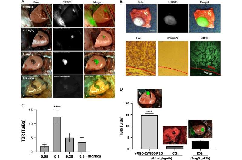
Therefore, the research teams developed dual-channel near-infrared fluorescence (cRGD-ZW800-PEG with 800 nm wavelengths, ZW700-1C with 700 nm wavelengths) and a technique of simultaneously exploring lung tumors and intersegmental lines using dual-channel fluorescence imaging during surgery. The feasibility of the dual-channel fluorescence imaging technique was evaluated in medium and large-sized animal models with lung cancer.
As a result, lung cancer and intersegmental lines were accurately explored simultaneously for up to 30 minutes through injection of cancer-targeting fluorescence (cRGD-ZW800-PEG) and contrast agents confirming blood flow distribution around cancer (ZW700-1C and ZW800-PEG) in the preclinical study (medium and large-sized animal models with lung cancer). This demonstrated the fluorescence agent’s high effectiveness during surgery.
In addition, dual-channel fluorescence is physically and chemically stable. Remarkably, this has excellent in vivo safety, as more than 85% of it is excreted through the kidneys within 4 hours after intravenous injection, which was confirmed by preclinical studies.

Prof. Kim, the corresponding author of the paper, said, “This study will open a new paradigm in image-guided cancer surgery via injection of cancer-targeting fluorescence (cRGD-ZW800-PEG) and ZW700-1C with excellent stability in vivo. Exploring lung tumors and intersegmental lines, which has been a difficult task so far, will now become easier using dual-channel fluorescence imaging technique.”
Prof. Hak Soo Choi emphasized the meaning of this study and said, “Dual-channel near-infrared fluorescence and imaging technique can be applied not only to lung cancer but to other cancers as well. By resecting only tumors, unnecessary resection of normal tissue will be minimized and patients will have improved quality of life.”
Prof. Kim also said, “This paper is the result of 5 years of continuous research by Korea University’s Guro Hospital and Harvard Medical School. We aim to focus on the clinical application of cancer-targeting fluorescence through additional research at research-oriented hospitals.”
More information:
Ok Hwa Jeon et al, Precise and safe pulmonary segmentectomy enabled by visualizing cancer margins with dual-channel near-infrared fluorescence, International Journal of Surgery (2024). DOI: 10.1097/JS9.0000000000001045
Provided by
Korea University College of Medicine
Citation:
Dual-channel fluorescence imaging for precise and safe pulmonary segmentectomy (2024, July 2)
retrieved 2 July 2024
from https://medicalxpress.com/news/2024-07-dual-channel-fluorescence-imaging-precise.html
This document is subject to copyright. Apart from any fair dealing for the purpose of private study or research, no
part may be reproduced without the written permission. The content is provided for information purposes only.

