A group of scientists at UCL have successfully created mechanical force sensors directly in the developing brains and spinal cords of chicken embryos, which they hope will improve understanding and prevention of birth malformations such as spina bifida.
The study, published in Nature Materials and in collaboration with the University of Padua and the Veneto Institute of Molecular Medicine (VIMM), uses innovative biotechnologies to measure the mechanical forces exerted by the embryo during its development.
These forces are crucial in the formation of organs and anatomical systems, such as the formation of the neural tube, which gives rise to the central nervous system.
Congenital spinal cord malformations affect around one in 2,000 newborns in Europe each year.
Although these malformations have been studied for decades, they cannot be fully explained through molecular and genetic studies alone.
As a result, researchers are now looking at the physical and mechanical forces in tissues during embryo development. However, this can be challenging as the embryonic spinal cord is tiny—too small to be seen with the naked eye—and extremely delicate. Force measuring devices therefore need to be similarly small and soft to avoid disrupting normal growth.
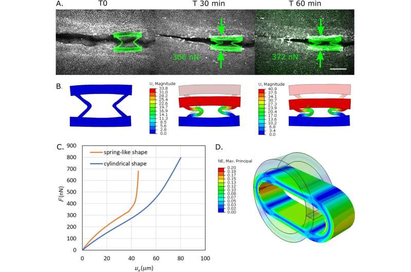
To overcome these difficulties, researchers 3D printed tiny force sensors (about 0.1mm wide) directly inside the developing nervous system of chicken embryos.
These force sensors begin as a liquid applied directly to the developing embryos. When exposed to a strong laser, the liquid transforms into a spring-like solid. This solid attaches to the growing spinal cord of the embryos and gets deformed by the mechanical forces produced by the embryo’s cells.
This enabled them to measure the minute forces—about a tenth of the weight of a human eyelash—that embryos must generate to form the spinal cord.

For normal embryonic development, these forces must be greater than the opposing negative forces.
Quantifying the forces would allow researchers to explore drugs capable of sufficiently increasing positive forces or decreasing negative ones, in order to help prevent congenital malformations such as spina bifida.
Such drugs could also complement the benefits of folic acid intake—a well-established strategy for preventing developmental problems before and during pregnancy.
Lead author, Marie Sklowdowska Curie postdoctoral fellow, Dr. Eirini Maniou (UCL Great Ormond Street Institute of Child Health and University of Padua), said, “Thanks to the use of novel biomaterials and advanced microscopy, this study promises a step change in the field of embryonic mechanics and lays the foundation for a unified understanding of development.
“Our work paves the way for identifying new preventative and therapeutic strategies for central nervous system malformations.”
The research group also demonstrated that the same technology could be applied to human stem cells, as they develop into spinal cord cells.
In the future, this would allow for comparisons between the stem cells of healthy donors and patients with spina bifida, with the goal of understanding why some people develop the condition.
Co-senior author, Dr. Gabriel Galea (UCL Great Ormond Street Institute of Child Health), said, “This technology is very versatile and widely applicable to many research fields, and we hope it will be quickly adopted and applied by other groups to address fundamental questions.”
Co-senior author, Professor Nicola Elvassore (University of Padua and VIMM), added, “This discovery not only allows us to better understand the mechanical forces at play during embryonic development but also offers new perspectives for intervening and preventing conditions like spina bifida.
“The ability to quantify embryonic forces with such precision represents a significant step forward in biomedical research.”
More information:
Eirini Maniou et al, Quantifying mechanical forces during vertebrate morphogenesis, Nature Materials (2024). DOI: 10.1038/s41563-024-01942-9
Citation:
New study used 3D-printed sensors to measure spinal cord malformations in embryos (2024, July 12)
retrieved 12 July 2024
from https://medicalxpress.com/news/2024-07-3d-sensors-spinal-cord-malformations.html
This document is subject to copyright. Apart from any fair dealing for the purpose of private study or research, no
part may be reproduced without the written permission. The content is provided for information purposes only.

