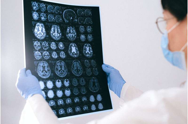
A research group at the Department of Biochemistry and Molecular Biology of the University of Seville has made a significant advance by discovering the crucial role of the protein Galectin-3 in the progression of various types of brain tumors.
In these tumors, the most abundant immune system cells, microglia and macrophages, overexpress Galectin-3, which creates an immunosuppressed environment that inhibits the action of other immune cells against cancer cells.
In vitro findings have shown that specific inhibition of Galectin-3 in microglial cells promotes expression of proinflammatory markers and reverses the presence of key immunosuppressive biomarkers. In vivo models of brain metastases of breast cancer and glioblastoma, two of the most common and aggressive types of brain tumors in the population, have validated these results.
In genetically modified models where Galectin-3 was knocked out, microglia cells and macrophages displayed a more inflammatory and anti-tumor state, leading to a reduction in the size of primary tumors and brain metastases by up to fivefold.
The research is published in the journal Cancer Letters.
These results, although preclinical, have clear translational potential. Thanks to collaborations with Lund University, the research group has access to the TD006 antibody, a selective inhibitor of Galectin-3 currently being tested in clinical trials in Alzheimer’s patients.
The team is currently working to improve Galectin-3 inhibitors so that they can improve their efficiency in reaching brain tumors, as well as researching their use in combination with other conventional therapies such as radiotherapy and chemotherapy.
More information:
Alberto Rivera-Ramos et al, Galectin-3 depletion tames pro-tumoural microglia and restrains cancer cells growth, Cancer Letters (2024). DOI: 10.1016/j.canlet.2024.216879
Citation:
The key role of Galectin-3 in brain tumor development (2024, May 3)
retrieved 4 May 2024
from https://medicalxpress.com/news/2024-05-key-role-galectin-brain-tumor.html
This document is subject to copyright. Apart from any fair dealing for the purpose of private study or research, no
part may be reproduced without the written permission. The content is provided for information purposes only.

